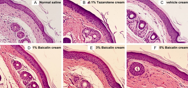
Figure 4.

Histological sections of mouse tail skin treated topically for 4 consecutive weeks and stained with HE (original magnification, × 200). Note: A. The granular layer is less developed in most parts of the mouse tail skin. B. A well-developed granular layer was clearly seen in mouse tail skin treated with 0.1% tazarotene cream. C-F. The degrees of orthokeratosis increased in a baicalin dose-dependent manner.
