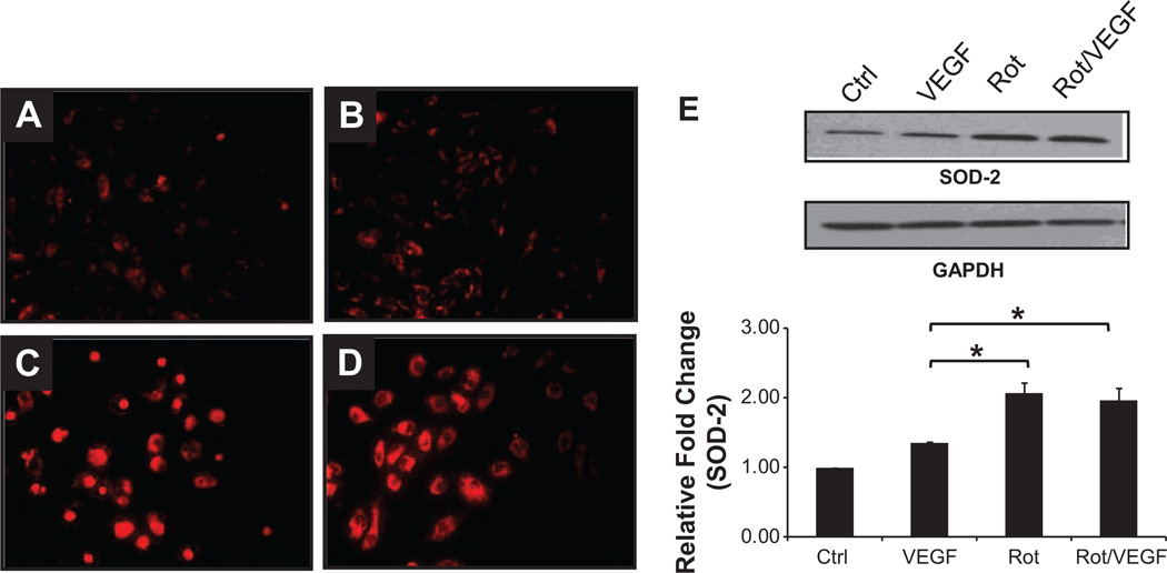
Figure 4.
Mitochondrial oxidative stress in human coronary artery endothelial cells (HCAECs). HCAECs were treated with vehicle (A), vascular endothelial growth factor (VEGF; 50 ng/mL; B), rotenone (Rot; 1 µmol/L; C), and Rot/VEGF (D). Rot increases mitochondrial superoxide production as shown by the higher fluorescence intensity of MitoSox Red (A and B vs C and D; n=3). Superoxide dismutase (SOD)-2 expression, normalized against GAPDH, was significantly increased in Rot-treated cells (E; n=3; *P<0.05), suggesting upregulation of this antioxidant as compensation to the elevated mitochondrial oxidative stress.

