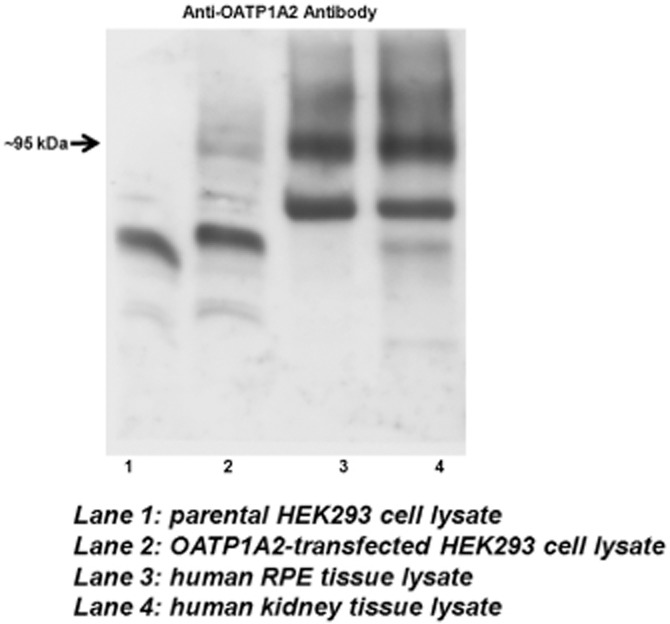
Figure 1.

Protein expression of OATP1A2 in human RPE by immunoblot analysis. Cells and human tissues were dissolved in RIPA buffer. Protein lysate was denatured at 55°C for 30 min and loaded onto SDS-PAGE. Protein signal was detected with anti-OATP1A2 antibody (∼95 kDa). Consistent findings were made in RPE tissue lysate obtained from four independent donors, with a representative image shown in this figure.
