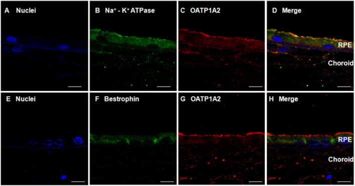
Figure 2.

Immunofluorescent labelling of OATP1A2 in human RPE. Sections of human retina were immunolabelled with anti-Na+-K+ ATPase β1 subunit antibody or anti-bestrophin antibody and Alexa Fluor® 488 conjugate donkey anti-mouse IgG or anti-OATP1A2 antibody and Alexa Fluor® 594 conjugate goat anti-rabbit IgG. Panel A shows nuclei (blue); panel B shows immunostaining of the Na+-K+ ATPase β1 subunit (green); panel C shows the immunostaining of OATP1A2 (red); panel D shows the merged images of panels A, B and C. Panel E shows nuclei (blue); panel F shows the immunostaining of bestrophin (green); panel G shows the immunostaining of OATP1A2 (red); panel H shows the merged images of panels E, F and G. Scale bars: 50 μm in panel A–H.
