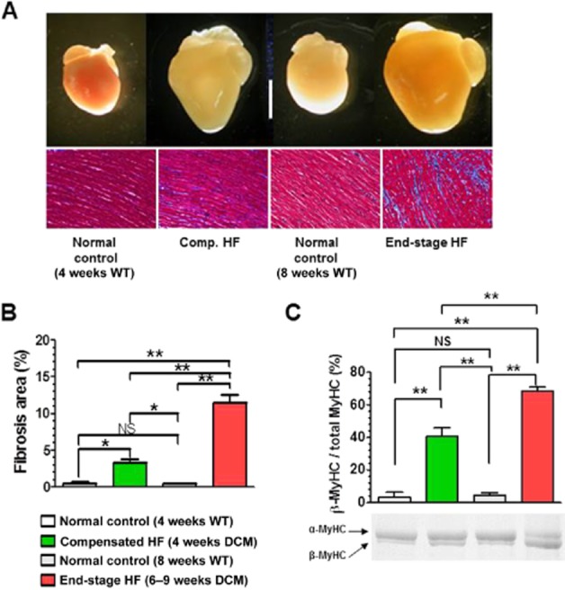
Figure 1.

Morphological and histological changes in the heart of DCM mice during progression of HF. (A) Gross morphology of the heart (top images) (scale bar, 5 mm) and histology of the LV (bottom images). Connective tissues were stained blue with Masson trichrome. (B) Quantitative analysis of the fibrosis area in the LV (n = 3 mice per group). (C) Protein expression level of MyHC isoforms in the LV (n = 5 mice per group). *P < 0.05; **P < 0.01; significantly different as indicated; one-way anova and post hoc Tukey's multiple comparison test.
