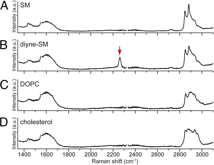
Fig. 2.
Raman spectra of supported lipid monolayers of (A) SM, (B) diyne-SM, (C) DOPC, and (D) chol on a quartz substrate. The Raman peak of diyne at 2,263 cm−1 is marked by a red arrow. The supported sample was prepared at 12 mN/m and 25 °C using the LB technique. Raman measurement was performed 15 times at different positions in each membrane, with an exposure time of 6 s. Averaged Raman spectra are shown. Raman peaks of O2 at ∼1,555 cm−1 and N2 at ∼2,330 cm−1 have been deleted so that Raman peaks from lipid molecules can be clearly seen, which is explained in Fig. S1.

