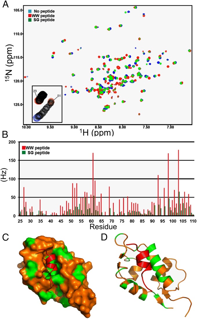
Fig. 5.
Interaction of MDMX WW and SG peptides with the p53-binding pocket. (A) Overlay of 1H-15N HSQC spectra for 15N-MDMX (blue), 15N-MDMX+WW peptide (red), and 15N-MDMX+SG peptide (green). (B) Chemical shift changes for MDMX p53BD residues binding to the WW peptide (red bars) or the SG peptide (green bars). (C) Surface image of the MDMX p53BD structure. The residues that have combined chemical shifts close to or greater than 50 Hz (upon binding the WW peptide) are highlighted in green. (D) Cartoon showing the chemical shifts on the back side of two helices obscured on the surface image.

