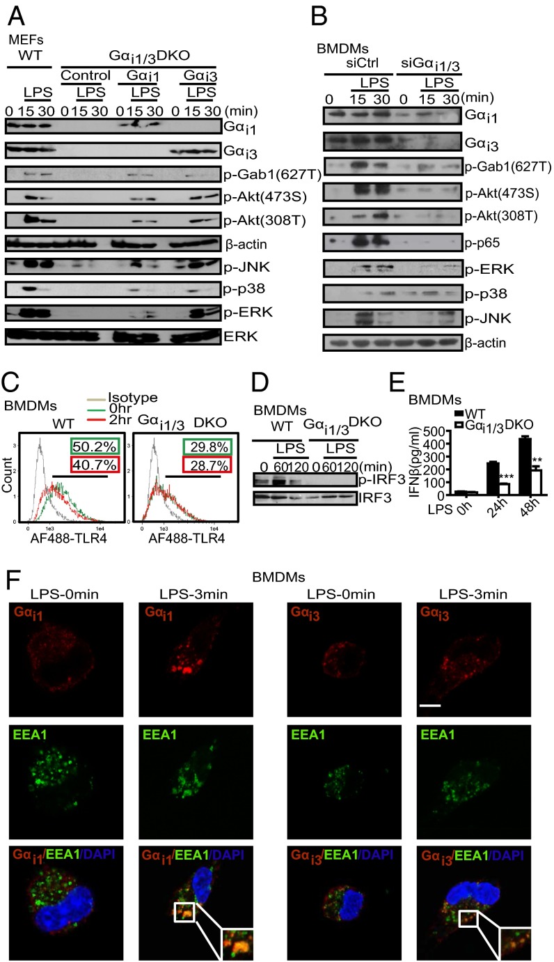
Fig. 2.
Gαi1/3 deficiency impairs the TLR4 triggered NF-κB, MAPKs, and TRIF signaling pathways. (A) Exogenous Gαi1 or Gαi3 rescue the activation of Akt and MAPKs signaling in DKO MEFs in response to LPS. DKO MEFs were transfected with vectors encoding WT Gαi1 and Gαi3 (4 μg per well). After 48 h, cells were treated with LPS (1 μg/mL) for the indicated times. Gαi1, Gαi3, p-Gab1 (T627), p-Akt (473S), p-Akt (308T), β-actin, p-JNK, p-p38, p-ERK, and total ERK were detected by Western blot. (B) Knockdown of the expression of Gαi1/3 decreases the activation of Akt and MAPKs by LPS in BMDMs. BMDMs were transfected with control small interfering RNA (siCtrl) or Gαi1 and Gαi3-specific siRNA. After 48 h, cells were stimulated with LPS (100 ng/mL) for the indicated times. Gαi1, Gαi3, p-Gab1 (627T), p-Akt (473S), p-Akt (308T), p-ERK, p-p38, p-JNK, and β-actin were detected by Western blot. (C) BMDMs from WT and Gαi1/3 DKO mice were treated or not with LPS (100 ng/mL) for 2 h. Flow cytometry was then used to examine TLR4 endocytosis by determining the surface levels of TLR4. (D) BMDMs from WT and Gαi1/3 DKO mice were treated or not with LPS (100 ng/mL) for the times indicated. The presence of phosphorylated IRF3 in cell extracts was determined. (E) BMDMs were treated with LPS (100 ng/mL) for the indicated times and the amounts of secreted IFN-β were determined by ELISA. (F) BMDMs were stimulated with LPS (100 ng/mL) for the times indicated and processed for confocal microscopy to detect the presence of Gαi1, Gαi3, or EEA1. (Scale bar: 5 μm.) All images are representative of at least three independent experiments where over 500 cells were examined per condition and >95% of the cells displayed similar staining.

