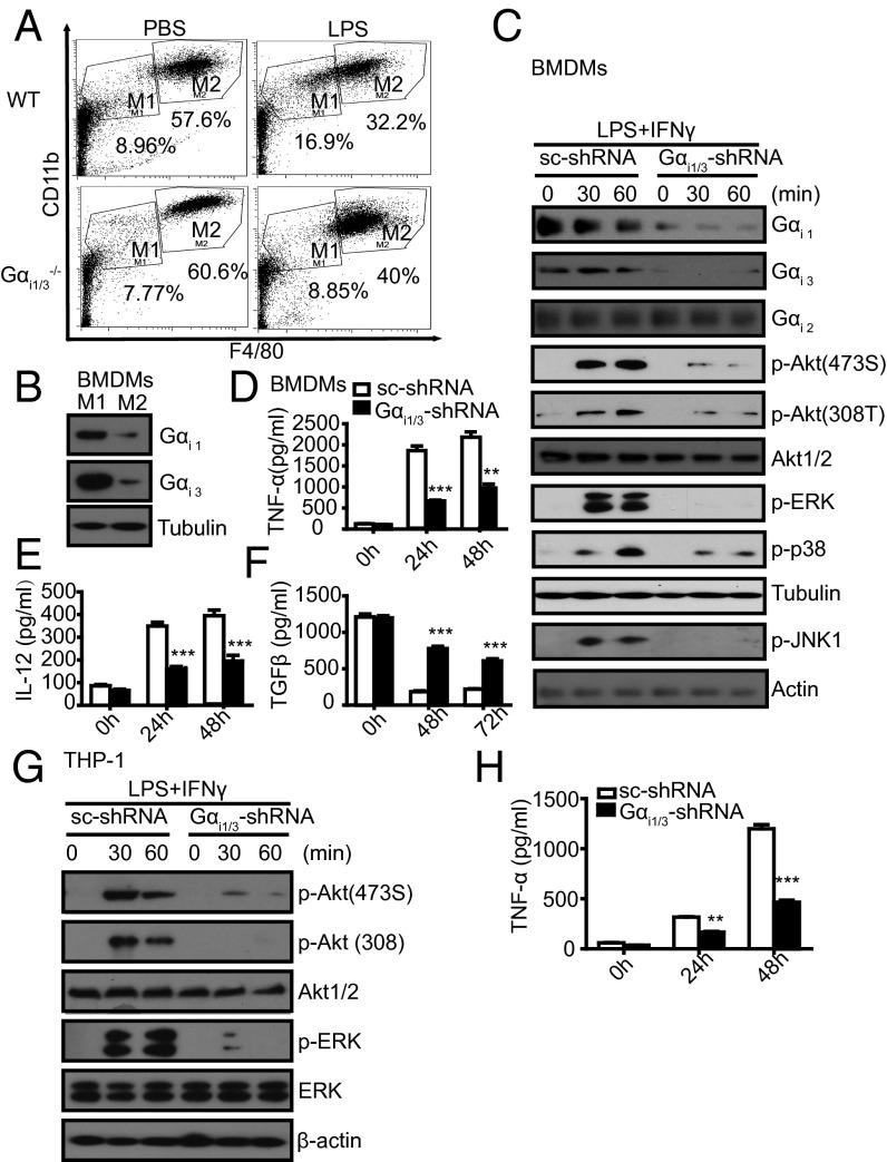
Fig. 4.
Gαi1/3 regulate macrophage polarization. (A) Peritoneal cells extracted from WT and Gαi1/3 DKO mice were analyzed by flow cytometry for CD11b and F4/80 to discriminate macrophage subtypes. WT or Gαi1/3 DKO mice were treated i.p. for 6 h with 20 mg/kg LPS or PBS. Peritoneal macrophages were extracted and subpopulations of macrophages were analyzed by flow cytometry with F4/80 and CD11b. Population M1 (F4/80intCD11bint) and population M2 (F4/80hiCD11bhi) were gated. (B) Gαi1/3 protein expression in M1 and M2 macrophages. BMDMs were treated with LPS (5 ng/mL) plus IFN-γ (10 U/mL) or IL-4 (10 U/mL), respectively, and tested for the M1 and M2 macrophage formation. After 24 h, cells were lysed. Gαi1 and Gαi3 were detected by Western blot. (C) Knockdown of Gαi1/3 decreased the activation of Akt and MAPKs in M1 macrophages. BMDMs were transfected with control small RNA (Ctrl) or Gαi1 and Gαi3-specific siRNA. After 48 h, BMDMs were treated with LPS (5 ng/mL) plus IFN-γ (10 U/mL) for the indicated times and were then lysed. Gαi1, Gαi2, Gαi3, p-Akt (473S), p-Akt (308T), total Akt, p-ERK1/2, p-p38, Tubulin, p-JNK, and β-actin were analyzed by Western blot. (D–F) Knockdown of Gαi1/3 decreases the expression of M1 cytokines: TNF-α (D), IL-12 (E), but increase the expression of the M2 cytokine TGF-β (F). BMDMs were transfected with control small RNA (Ctrl) or Gαi1 and Gαi3-specific siRNA. After 48 h, BMDMs were treated with LPS (5 ng/mL) plus IFN-γ (10 U/mL) for the indicated times, and TNF-α, IL-12, and TGF-β in the supernatants were measured by ELISA. **P < 0.01, ***P < 0.001. (G) Knockdown of Gαi1/3 decreases the activation of Akt and ERK in the THP-1 cell line. THP-1 cells were treated with LPS (5 ng/mL) plus IFN-γ (10 U/mL) for the indicated times and were then lysed. P-Akt (473S), p-Akt (308T), total Akt, p-ERK, total ERK, and β-actin were analyzed by Western blot. (H) Knockdown of Gαi1/3 decreases the expression of the M1 cytokine TNF-α in THP-1 cells. THP-1 cells were transfected with control small interfering RNA (Ctrl) or Gαi1 and Gαi3-specific siRNA. The level of TNF-α in the supernatants was measured by ELISA. **P < 0.01, ***P < 0.001.

