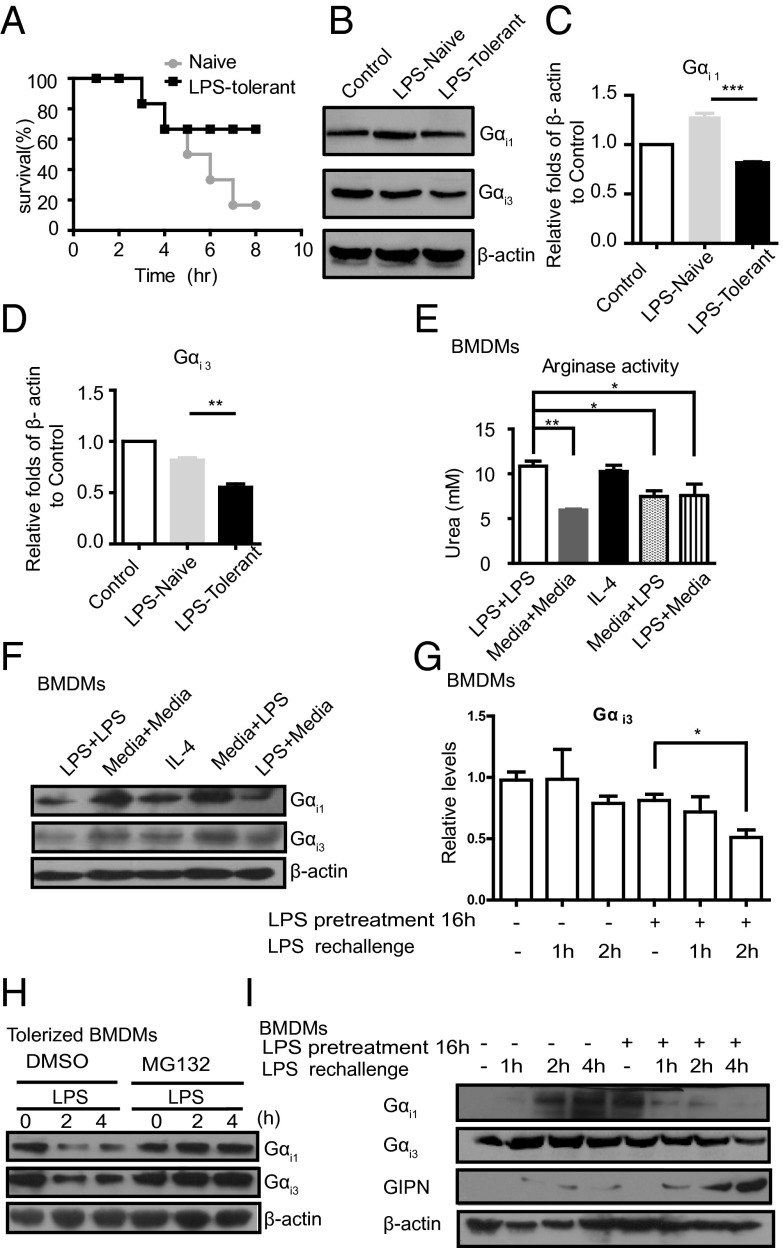
Fig. 5.
Gαi1/3 degradation is involved in LPS tolerance. (A) Survival of mice (n = 6 per group) given saline (Naive) or preexposure to a low dose of LPS (LPS-tolerant) were challenged with a lethal dose of LPS (4 mg/kg. i.p.) plus d-galactosamine (500 mg/kg). Survival was monitored over 10 h. (B) Gαi1/3 expression in peritoneal macrophages (primary Mφ) isolated from mice treated as in A for 2 h. Gαi1, Gαi3 was detected by Western blot. (C and D) Expression of Gαi1/3 level was quantified from Western blot assays. The data are expressed as the mean ± SE of the ratios of indicated protein to β-actin. **P < 0.01, ***P < 0.001. (E) BMDMs (2 × 10⁵ cells per well) were tolerized or not with 100 ng/mL LPS for 16 h, washed, and rechallenged with 100 ng/mL LPS. After 8 h of incubation, arginase activity was assessed by an assay of urea production from arginine substrate. *P < 0.05, **P < 0.01. (F) Expression of Gαi protein in BMDMs tolerized or not with 100 ng/mL LPS for 16 h, washed, and rechallenged with 100 ng/mL LPS. After 8 h of incubation, cells were lysed. Gαi1, Gαi3, and β-actin were assessed by Western blot. (G) BMDMs were vehicle-treated or tolerized with 100 ng/mL LPS for 16 h, washed, and rechallenged with 100 ng/mL LPS for the indicated times. Gαi3 mRNA expression was measured by QRT-PCR. *P < 0.05, **P < 0.01. (H) BMDMs tolerized overnight with LPS (100 ng/mL) were pretreated for 30 min with either vehicle only (DMSO) or MG132 (25 μM). Cells were subsequently stimulated with LPS (100 ng/mL) for the indicated times and lysed. Gαi1, Gαi3, and β-actin were assessed by Western blot. (I) BMDMs were vehicle-treated or tolerized with 100 ng/mL LPS for 16 h, washed, and rechallenged with 100 ng/mL LPS for indicated times. Gαi1, Gαi3, GIPN, and β-actin were assessed by Western blot.

