Fig. 1.
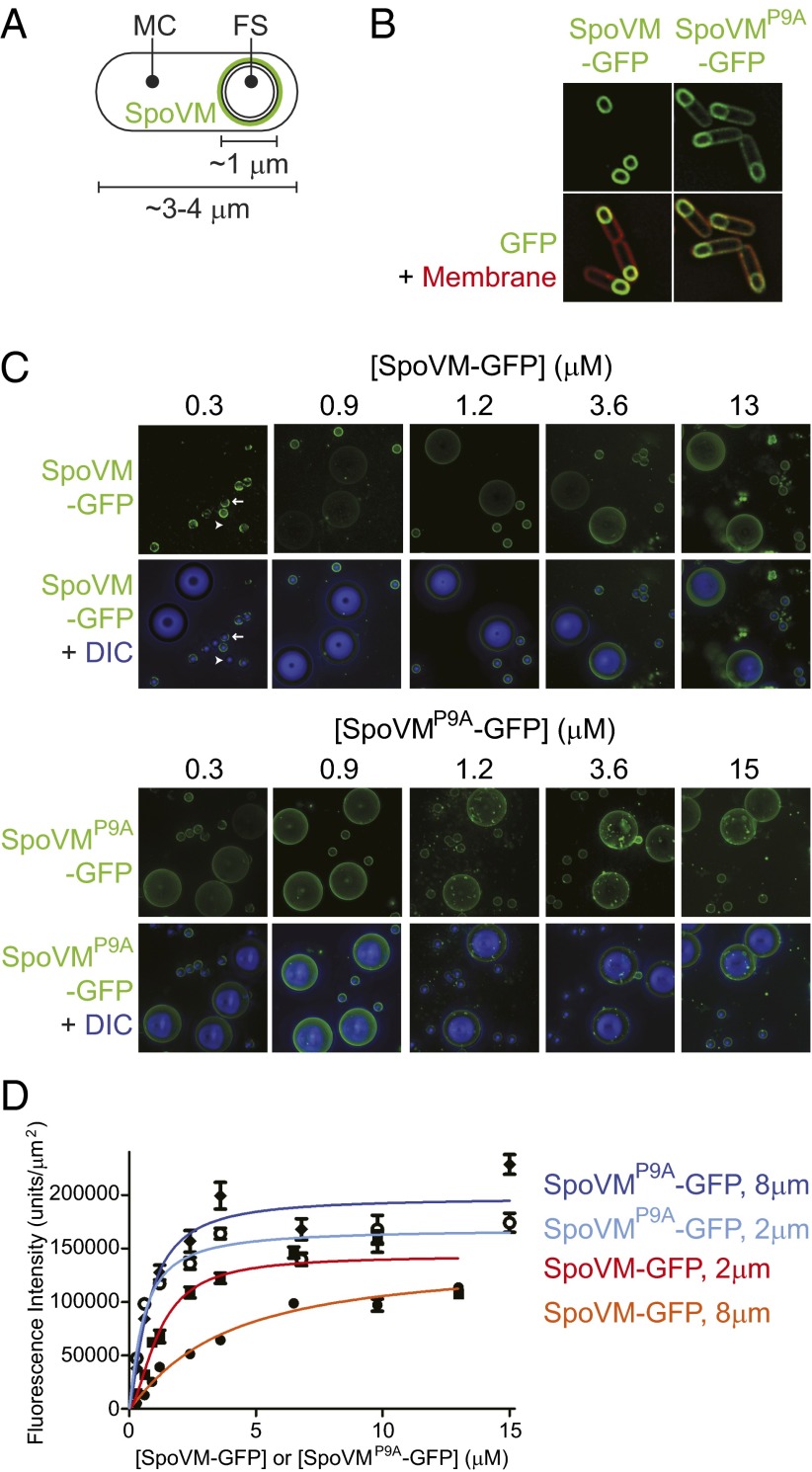
SpoVM, but not SpoVMP9A, preferentially localizes to convex membranes. (A) Schematic of a sporulating B. subtilis cell, in which the rod-shaped mother cell (MC) elaborates a spherical internal organelle termed the forespore (FS). SpoVM (green) is produced exclusively in the MC and localizes to the FS surface. Membranes are depicted as black lines. (B) Fluorescence micrographs of sporulating B. subtilis cells. In vivo localization of SpoVM-GFP (Left) or SpoVMP9A-GFP (Right). Overlay of GFP signal (green) and membranes (red) visualized using the fluorescent dye FM4-64. (C) Fluorescence micrographs of SSLBs 2 or 8 μm in diameter that were incubated with increasing concentrations of purified SpoVM-GFP (Upper) or SpoVMP9A-GFP (Lower). Aggregate GFP fluorescence across several z-stacks (top of each data set) is depicted in green; the bottom of each data set shows the overlay of GFP fluorescence (green) and SSLBs visualized by differential interference contrast (DIC) microscopy (blue). Arrow: bead displaying a nonuniform pattern of fluorescence; arrowhead: bead displaying little or no fluorescence (see Fig. S1 for magnified view). (D) Quantification of SpoVM-GFP fluorescence on 2-μm (red) or 8-μm (orange) SSLBs or SpoVMP9A-GFP on 2-μm (light blue) or 8-μm (blue) SSLBs per square micron, as a function of protein concentration. Each data point represents the mean intensity of 30–52 SSLBs; errors bars are SEM; data were fit with an allosteric sigmoidal model.

