Fig. 5.
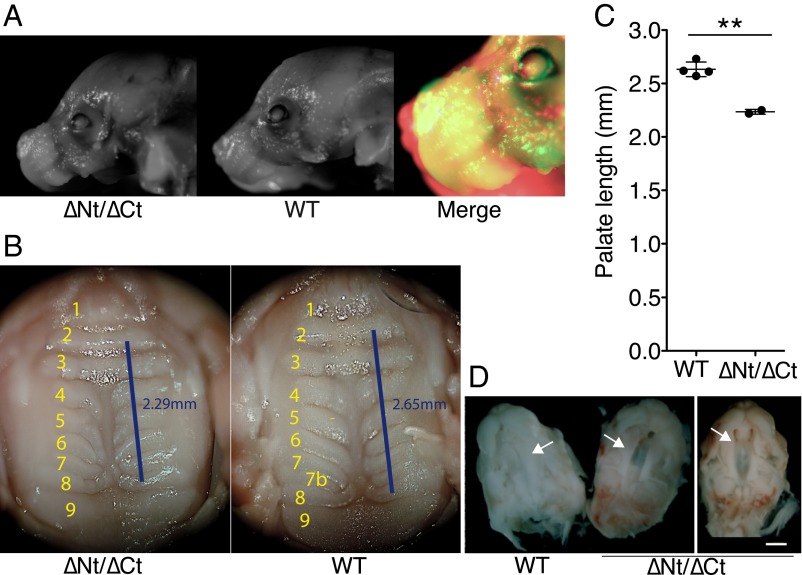
Craniofacial defects in didoΔNT/ΔCT neonates. (A) didoΔNT/ΔCT mice showed altered craniofacial development at birth, with a shortened snout compared with WT littermates. Representative images of both genotypes are shown, with a false-color merged image for comparison. (B and C) Palate was fully closed at birth, but the distance between rugae 2 and 8 was 15% shorter in didoΔNT/ΔCT than in WT pups (WT, n = 4; didoΔNT/ΔCT, n = 2; Student's t test P = 0.0016). (D) Delayed palate closure in didoΔNT/ΔCT embryos. At 15.5 dpc, embryos were extracted and the lower jaw and tongue removed to expose the developing palate. The secondary palate was fully closed in WT embryos (Left), whereas didoΔNT/ΔCT embryos showed varying degrees of delay in palate closure, with partial (Center) or almost no (Right) horizontal growth of palatal halves (arrows) toward the midline at the time analyzed (n = 8). (White scale bar, 5 mm.)

