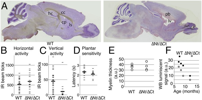
Fig. 6.
Neural anomalies in dido mutant mice. (A) Sagittal sections of brain from a representative didoΔNT/ΔCT mouse, showing ventricle enlargement and dysplasia of corpus callosum (cc) and hippocampus (hp). cp, caudoputamen; ls, lateral striatum; lv, lateral ventriculum; Pb, Probst bundles. (B) Horizontal and (C) vertical spontaneous locomotor activity in surviving adult didoΔNT/ΔCT mice. Each mouse was tested three times over a 4-mo period. One-tailed Student's t test, P = 0.04 for vertical differences. (D) Plantar sensitivity to thermal stimulus in surviving adult didoΔNT/ΔCT mice. (B–D) As one WT and two didoΔNT/ΔCT mice died during this period, two values for n are given for each group; n = 10 (9) for WT, n = 7 (5) for didoΔNT/ΔCT mice. (E) Toluidine-stained myelin was photographed in cross-sections of left and right sciatic nerves from adult didoΔNT/ΔCT mice and WT littermates, and myelin sheet thickness was measured with Adobe Photoshop (n = 6 for WT, n = 4 for didoΔNT/ΔCT); 20 axons per nerve were evaluated in a blind manner. (F) Peripheral nerve α-tubulin deacetylation in didoΔNT/ΔCT and WT mice (n = 4 + 4). Optical density of acetylated tubulin bands in Western blots of sciatic nerve extracts, normalized to total α- + β-tubulin levels. One-tailed paired Student's t test; P = 0.02.

