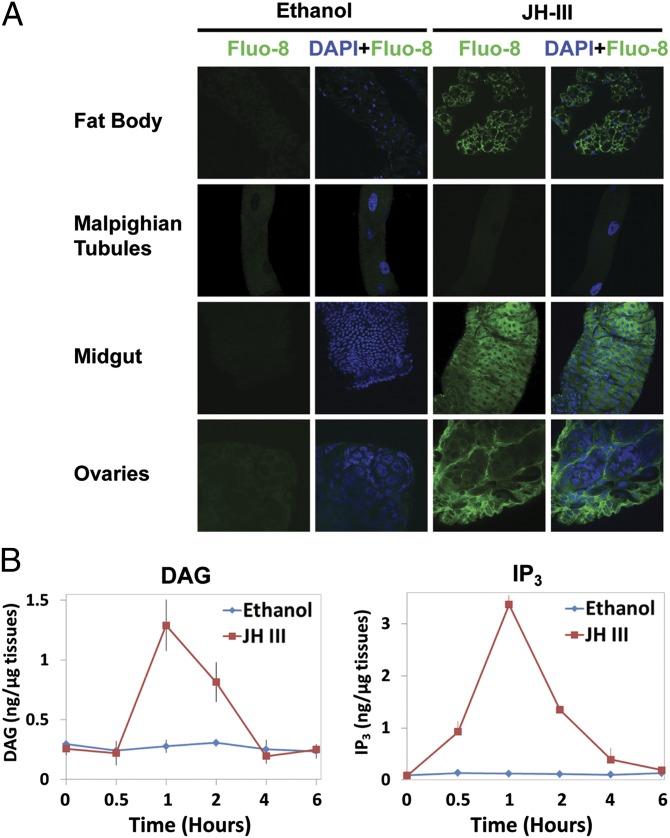
Fig. 2.
JH treatment causes a rise in intracellular levels of calcium, DAG, and IP3 in mosquito tissues. (A) JH-stimulated calcium responses in the fat body, Malpighian tubules, midgut, and ovaries. Tissues dissected from adult female mosquitoes within 30 min PE were preincubated with Fluo-8 AM in APS for 1 h, followed by incubation with 1 μM JH-III or ethanol for 15 min. Images were captured using a Zeiss LSM 510 confocal microscope at 1,000× magnification. Cell nuclei were stained blue with DAPI, whereas the binding of calcium to Fluo-8 AM greatly enhanced the green fluorescence intensity. Representative images were taken from one of three independent experiments with similar results. (B) JH treatment increased the production of DAG and IP3 in cultured fat bodies that were isolated from newly emerged mosquitoes. Fat bodies were incubated in the tissue culture medium with JH-III at a final concentration of 1 µM. Ethanol was used as a negative control. The experiment was repeated twice with three replicates per treatment.

