Fig. 3.
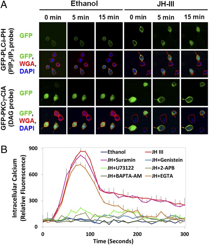
JH activates the phospholipase C–calcium pathway. (A) The amount of PIP2 at the plasma membrane decreased in Aag2 cells after exposure to JH-III. Aag2 cells were transfected with plasmids encoding GFP-PLCδ-PH or GFP-PKCγ-C1A and were stained with DAPI (blue) and the plasma membrane marker WGA (red). The PH domain of PLCδ binds with high affinity to PIP2 and IP3. The C1A domain of PKCγ has a high affinity for DAG. Subcellular translocation of the GFP reporters after JH treatment was captured using a confocal microscope at 1,000× magnification. Representative images are shown. (B) Intracellular calcium was quantitated after Aag2 cells were incubated with 1 µM of JH-III and the indicated chemicals that blocked the PLC–Ca2+ signaling pathway. Ethanol was used as a negative control. Data are presented as mean ± SD (n = 3).

