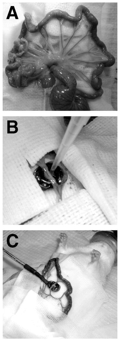
FIG. 2.

Gene transfer and electroporation of the mesenteric vasculature. (A) The mesenteric vascular tree. (B) DNA solution being pipetted into the electrode. The vessel is draped into the bath electrode, which is surrounded by surgical drapes to maintain a sterile field. At the end of the electroporation of this vessel, the DNA solution can be removed and reused for the next vessel. (C) Placement of electrode during surgery.
