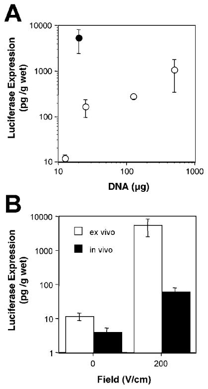
FIG. 3.
Comparison of gene expression in vivo and ex vivo following gene transfer to the lungs using electroporation. (A) Dose–response curve for ex vivo gene transfer in mice and rats. pCMV-lux-DTS plasmid at the indicated doses was administered to excised mouse (closed circles: female Balb/c, 15–20 g) or rat (open circles: male Sprague–Dawley, 150–400 g) lungs via the bronchi and the individual lobes were electroported by direct placement of electrodes on the lobes using eight square wave pulses of 10 msec duration each at 200 V/cm. Mouse lungs received 200 μl of plasmid and rat lungs received 500 μl. Lungs were placed into medium and luciferase gene expression was measured 24 h later (mean ± SEM, n = 3). (B) Comparison of in vivo and ex vivo gene transfer and expression in the mouse lung. Ex vivo transfer was performed as described in (A). For in vivo electroporation, 20 μg of pCMV-Lux-DTS in 100 μl were injected intratracheally and varying field strengths were applied to the chest of mice (eight pulses of 10-msec duration each). The levels of luciferase gene expression were measured at 2 days posttreatment as previously described (Dean et al., 2003) (mean ± SEM; n = 4 animals/point). Values from (A) are shown for ex vivo expression at the same DNA dose (Dean et al., 2003).

