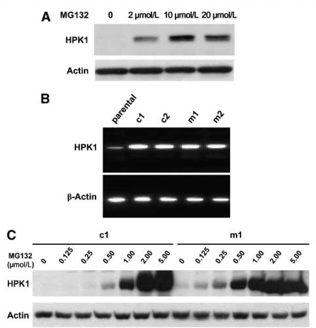
Figure 3.
HPK1 proteins were targeted by proteasome-mediated degradation in PDA cell lines. A, Western blots showing that MG132 treatment enhanced HPK1 protein expression in a dose-dependent manner in Panc-1 cells.Panc-1 cells were treated with 2.0 to 20.0 µmol/L of MG132 for 16 h. B, RT-PCR results showing HPK1 mRNA expression in Panc-1 parental cells, wild-type HPK1/ Panc-1 stable clones (c1 and c2), and kinase-dead mutant HPK1/Panc-1 stable clones (m1 and m2). C, Western blots showing that MG132 treatment (0.125–5.0 µmol/L for 16 h) enhanced HPK1 protein levels in a dose-dependent manner in c1 and m1 stable cells. Actin was used as the loading control.

