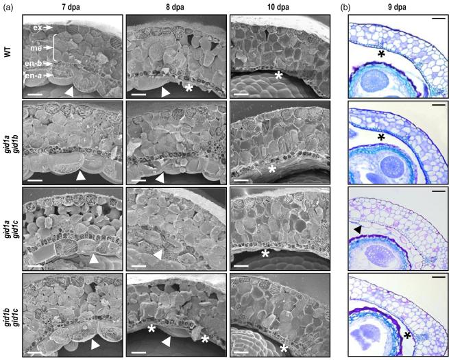
Figure 6.
The gid1 null mutants show morphological alterations in fruit structure.
(a) Delayed degradation of endocarp a in double gid1a gid1b and gid1a gid1c mutants. Transverse cryosections of fruits at 7, 8 and 10 dpa of wild-type (WT) and double mutants are shown. In the left top image, the different tissues are labeled: ex, exocarp; me, mesocarp; en-b, endocarp b; en-a, endocarp a. Presence or degradation of en-a are indicated by an arrowhead or asterisk, respectively. Scale bar represents 50 μm.
(b) Delayed lignification of en-b and degradation of en-a in double gid1a gid1c mutant. Transversal sections of fruits at 9 dpa of WT and the double gid1 mutants are shown. Presence of end-a and delayed lignification of end-b in gid1a gid1c mutant is indicated by an arrow head; degradation of en-a and strong lignification of en-b in WT and in gid1a gid1b and gid1b gid1c mutant is indicated by an asterisk. Scale bar represents 50 μm.

