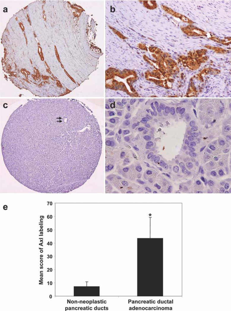
Figure 1.
Representative micrographs show (a and b) strong cytoplasmic staining for Axl in a moderately differentiated PDA and (c and d, arrows) no expression of Axl protein in normal pancreatic duct (original magnification, 20× for a and c, 200× for b, and 400× for d). (e) Axl expression in PDA samples and their paired non-neoplastic pancreatic ductal epithelial cells are shown. *P < .01.

