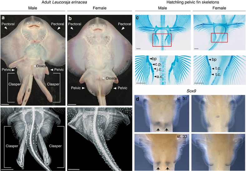
Figure 1. Morphology and development of L. erinacea claspers.
(a,b) Ventral view of sexually mature male (a) and female (b) skates. In these panels the scale bar, 2.5 cm. Lower panels show corresponding X-rays of the male and female pelvic fins with scale bars, 4 cm. (c) Alcian blue staining of hatchling pelvic fins. The posterior skeletal elements of males and females are labelled as follows: bp, basipterygium; t.c., terminal cartilages; c.p., covering plate; j.c., junctional cartilages; a.c., axial cartilage. Scale bars, 1 mm. (d) Sox9 expression in developing male (left) and female (right) pelvic fins at stages 31 and 33.

