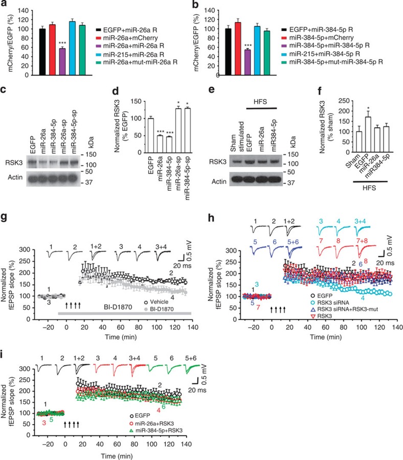
Figure 3. RSK3 mediates the effect of miR-26a and miR-384-5p on LTP.
(a,b) Cultured hippocampal neurons were transfected with designated reporter constructs at DIV14 and fixed for image acquisition at DIV17; n=13–15 neurons for each group; data are presented as mean±s.e.m.; Kruskal–Wallis and Mann–Whitney U-tests are used for statistical analysis; ***P<0.001; scale bar, 20 μm. (c–f) Cultured hippocampal slices were transduced with a designated lentivirus, unstimulated (c,d), sham-stimulated (e,f) or stimulated with high-frequency stimulation (HFS; e,f) and immunoblotted for RSK3; data are presented as mean±s.e.m.; n=4–5 slices for each condition; Kruskal–Wallis and Mann–Whitney U-tests are used for statistical analysis; *P<0.05, ***P<0.001. (g–i) Cultured hippocampal slices were treated with vehicle or BI-D1870 or transduced with designated lentivirus, and stimulated for LTP induction; the fEPSP slope normalized to the baseline prior to stimulation was plotted as mean±s.e.m.; n=5–7 slices for each condition; one-way analysis of variance and Student's t-test are used for normally distributed data with equal variance, and Kruskal–Wallis and Mann–Whitney U-tests are used for non-normally distributed data with unequal variance for statistical analysis.

