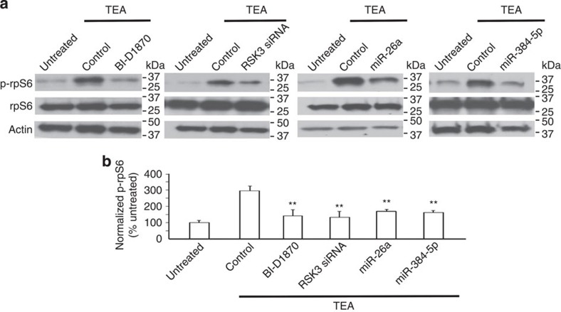
Figure 4. Increased rpS6 phosphorylation caused by the expression change to miR-26a, miR-384-5p and RSK3 in LTP.
Primary cortical neurons (DIV4) were transduced with lentivirus-expressing EGFP (as the control), RSK3 siRNA, miR-26a or miR-384-5p. At 2 weeks after transduction, neurons were treated with TEA (25 mM, 15 min) and harvested at 90 min after treatment for immunoblotting. BI-D1870 was added to neural medium at 5 min before TEA treatment. (a) Representative blots. (b) Quantification of a. Data are presented as mean±s.e.m. n=4–5 experiments for each condition. Kruskal–Wallis and Mann–Whitney U-tests are used for statistical analysis. **P<0.01.

