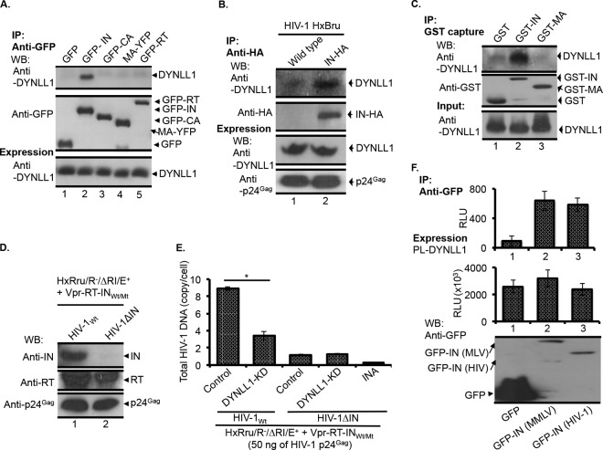
FIG 2.
HIV-1 IN, but not CA, MA, or RT, interacts with DYNLL1. (A) The GFP, GFP-IN, GFP-CA, MA-YFP, or GFP-RT protein was expressed in 293T cells, and coprecipitation of endogenous DYNLL1 (top panel) was detected by Co-IP using an anti-GFP antibody, as described in Materials and Methods. The immunoprecipitation of GFP, GFP-IN, MA-YFP, or GFP-CA (middle panel) was detected by WB using an anti-GFP antibody. DYNLL1 expression in the total cell lysate was detected by WB using an anti-DYNLL1 antibody (bottom panel). The data were confirmed in three independent experiments. (B) IN-DYNLL1 interaction during HIV-1 infection. C8166T cells (15 × 106) were infected with the HxBru or HxBru-IN-HA virus (at 50 ng of virus-associated p24Gag). After 72 h p.i., the cells were subjected to Co-IP using an anti-HA antibody, and coprecipitation of DYNLL1 (top panel) was detected by WB using an anti-DYNLL1 antibody, as described in Materials and Methods. IN-HA immunoprecipitation was determined by probing the WB with an anti-HA antibody (second panel from the top). The expression of DYNLL1 and similar level of HIV-1 infection were determined by probing the total cell lysates for the DYNLL1 and HIV-1 p24Gag proteins by WB (third and fourth panels from the top, respectively) using the corresponding antibodies. The data were confirmed in two independent experiments. (C) IN-DYNLL1 interaction in vitro. GST, GST-IN, or GST-MA protein was incubated with recombinant DYNLL1 protein. The coprecipitation of DYNLL1 (top panel) was detected as described in Materials and Methods. The immunoprecipitation of GST, GST-IN, or GST-MA in elutes was detected by WB using an anti-GST antibody (middle panel). DYNLL1 protein in the supernatants was detected by WB using an anti-DYNLL1 antibody (bottom panel). The data were confirmed in two independent experiments. (D) HIV-1Wt or HIV-1ΔIN virus-incorporated RT (top panel), IN (middle panel), and p24Gag (bottom panel) was detected by WB using the corresponding antibodies, as described in Materials and Methods. (E) Control or DYNLL1-KD C8166T cells (1.5 × 10⁶) were infected with the HIV-1Wt or HIV-1ΔIN virus (at 50 ng of virus-associated p24Gag). At 12 h p.i., total HIV-1 DNA was quantified by qPCR. The values shown are the averages of triplicates with the indicated standard deviations. The data were confirmed in two independent experiments. Statistical significance was determined using Student's t test. *, P < 0.05 (n = 3). (F) DYNLL1 interaction with MMLV IN. GFP, GFP-IN (MMLV), or GFP-IN (HIV-1) was cotransfected with PL-DYNLL1 in 293T cells. After 48 h of transfection, 8/10 of the cells were subjected to Co-IP with the anti-GFP antibody. The coprecipitation of PL-DYNLL1 was detected by measuring the PL activity from the immunoprecipitates (top panel). One-tenth of the cells were subjected to PL-DYNLL1 expression analysis by measuring the PL activity (middle panel). One-tenth of the cells were subjected to GFP, GFP-IN (MMLV), or GFP-IN (HIV-1) expression analysis by WB using the anti-GFP antibody (bottom panel). RLU, relative light units. INA, heat-inactivated virus control.

