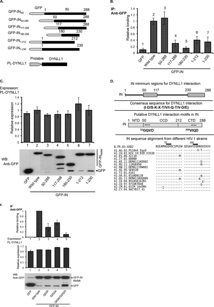
FIG 3.
The consensus motifs in the N- and C-terminal domains of IN are required for DYNLL1 interaction. (A) Schematic diagram of the GFP-IN deletion mutant and PL-DYNLL1 fusion proteins that were used in this study. (B and C) The GFP and GFP-INWt/deletion mutant proteins were coexpressed with PL-DYNLL1 in 293T cells. (B) Coprecipitation of PL-DYNLL1 was detected by Co-IP using an anti-GFP antibody, followed by analysis of PL activity from the immunoprecipitates, as described in Materials and Methods. (C) Upper panel, PL-DYNLL1 expression was detected by measuring PL activity from total cell lysate. The data are presented as fold change in the PL activity with respect to the wild-type control. Lower panel, the expression of GFP-INWt/Mt proteins in the total cell lysate was detected by WB using an anti-GFP antibody. The data shown are the average values from three independent experiments with the indicated standard deviation. (D) Diagram showing the minimum regions in HIV-1IN (i.e., aa 50 to 117 and 230 to 288) for DYNLL1 interaction, the general consensus sequence for DYNLL1 interaction, the putative DYNLL1 interaction motifs in HIV-1IN, and the IN sequence alignment for few representative HIV-1 strains. The full list of HIV-1 sequences, including the accession numbers, can be found in the HIV sequence compendium, 2014. (The IN sequence alignment is adapted from the HIV sequence compendium, 2014.) (E) The GFP-INWt, GFP-INQ53A, GFP-INQ252A, or GFP-INQ53A/Q252A protein was coexpressed with PL-DYNLL1 in 293T cells, and coprecipitation of PL-DYNLL1 (top panels) was detected by Co-IP using an anti-GFP antibody, followed by analysis of PL activity from the immunoprecipitates. PL-DYNLL1 expression was detected by measuring PL activity from total cell lysate (middle panel). The data are presented as fold change in the PL activity with respect to wild-type control. The expression of GFP-INWt/Mt (bottom panel) was detected by WB using an anti-GFP antibody. The data shown are the average values from three independent experiments with the indicated standard deviations.

