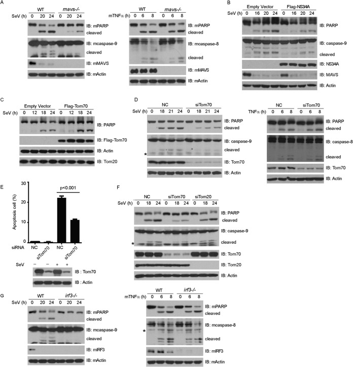
FIG 3.
MVAS, Tom70, and IRF3 mediate Sendai virus-induced apoptosis. (A) Wild-type (WT) and mavs−/− MEF cells were infected with SeV (MOI = 3) (left) or treated with TNF-α (10 ng/ml) and CHX (10 ng/ml) (right) for the indicated times. Cell lysates were collected for Western blot analysis with the indicated antibodies. (B and C) HEK293 cells were transfected with empty vector, Flag-NS3/4A (B), or Flag-Tom70 (C) for 24 h and then infected with SeV for the indicated times. Cell lysates were collected for Western blot analysis. (D) HEK293 cells were transfected with NC or Tom70 siRNA and then infected with SeV (left) or treated with TNF-α (10 ng/ml) and CHX (10 ng/ml) (right) for the indicated times. Cell lysates were collected for Western blot analysis with the indicated antibodies. (E and F) HEK293 cells were transfected with the indicated siRNAs and infected with SeV. (E) Cell apoptosis was quantified using annexin V staining and flow cytometry. (F) Cell lysates were collected for Western blot analysis with the indicated antibodies. (G) Wild-type (WT) and irf3−/− MEF cells were infected with SeV (MOI = 3) (left) or treated with mTNF-α (10 ng/ml) and CHX (10 ng/ml) (right) for the indicated times. Cell lysates were collected for Western blot analysis with the indicated antibodies. NC, negative-control siRNA; *, nonspecific band.

