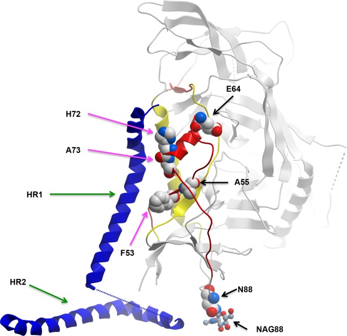
FIG 3.

Location of the T/F signature residues on the gp120-gp41 interactive region. The figure depicts one monomer of gp120 (gray) and gp41 (dark blue helix) from 4NCO (39). Mobile layer 1 is shown in red, and mobile layer 2 is shown in yellow. The mobile layers were identified in references 3 and 40. The black arrows point toward the T/F residues, and the magenta arrows point toward the gp41 contact residues identified (39). The numbers are HXB2 coordinates. The green arrows point toward heptad-repeat 1 (HR1) and heptad-repeat 2 (HR2) of gp41.
