
FIG 6.
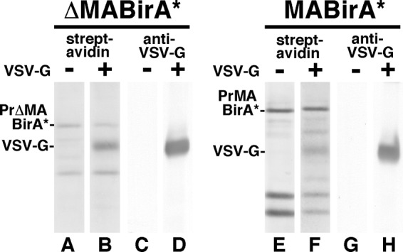
Detection of biotinylated VSV G protein in virus particles. ΔMA-BirA* (lanes A to D) and MA-BirA* (lanes E to H) constructs were transfected alone (lanes A, C, E, and G) or cotransfected with a VSV G protein expression construct (lanes B, D, F, and H). Three days posttransfection, proteins from virus particles were separated electrophoretically and blotted to detect biotinylated proteins (lanes A and B and lanes E and F) or the VSV G protein (lanes C and D and lanes G and H). PrΔMABirA*, PrMABirA*, and VSV G bands are as indicated.