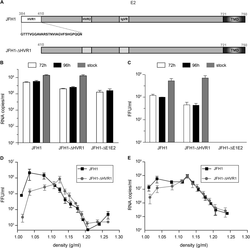
FIG 1.
(A) Schematic representation of the HVR1 deletion in E2. TMD, transmembrane domain. (B to E) Characterization of the JFH1-ΔHVR1 mutant. Huh-7 cells were electroporated with in vitro-transcribed JFH1 or JFH1-ΔHVR1 RNA, and cell supernatants were collected either 72 h or 96 h later. Viral stocks were produced after further amplification. Amplified virus stocks were precipitated and loaded on a 10 to 50% iodixanol gradient. The HCV genomes were quantified by quantitative RT-PCR (B), and HCV titers were determined (C). Results are expressed as the means from three independent experiments, and error bars represent the standard deviations of the means. Amplified virus stocks were precipitated and loaded on a 10 to 50% iodixanol gradient. After ultracentrifugation, 12 fractions were collected and the HCV genome copies (D) and titers (E) were quantified for each fraction. Results are expressed as the means from three independent experiments, and error bars represent the standard deviations of the means.

