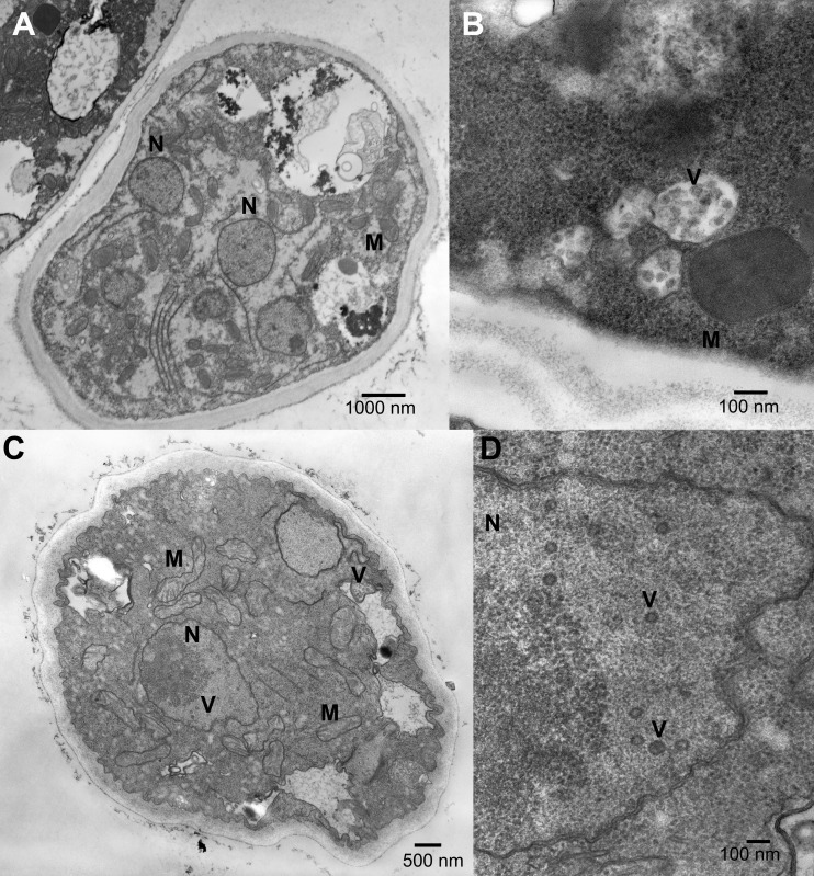
FIG 5.
Transmission electron micrographs of thin sections of Sclerotinia sclerotiorum isolates 328 and DK3 showing the degradation of mitochondria (M) and of nuclei (N) and the formation of lipid vesicles (V) associated with mycovirus infection. Cross sections through mycelia of virus-free S. sclerotiorum isolate DK3 (A), isolate DK3 transfected with SsHV2L showing vesicles associated with cell walls (B), and isolate 328 infected with SsHV2L and SsEV1 (C) and a high-magnification image of isolate 328 infected with SsHV2L and SsEV1 showing vesicles associated with the nucleus and cytoplasm (D).

