FIG 2.
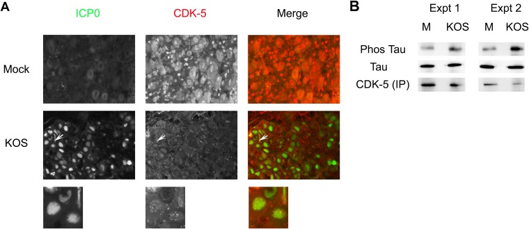
HSV-1 affects CDK-5 localization and kinase activity. (A) CDK-5 localization in response to HSV-1 infection. CD-1 mice were infected with 2 × 10⁵ PFU of KOS per eye. Three days postinfection, mice were sacrificed, and TG were collected, fixed, paraffin embedded, and processed for immunofluorescence staining of ICP0 and CDK-5. More than 10 sections from two independent experiments were examined, and the image shown is a representative section. Magnification, 400×. Arrows point to the expanded view shown below each panel. In about 30% of the ICP0-positive neurons, CDK-5 colocalized with ICP0 in the nucleus but showed punctate staining, as evident in the expanded view. (B) In vitro CDK-5 kinase assay. SK-N-SH cells were mock infected or infected with KOS for 24 h. CDK-5 protein was immunoprecipitated (IP) and incubated with bacterially purified Tau protein. Phosphorylated (Phos) versus total Tau was determined by Western blot analysis and quantified by densitometry. Data from two independent experiments are shown.

