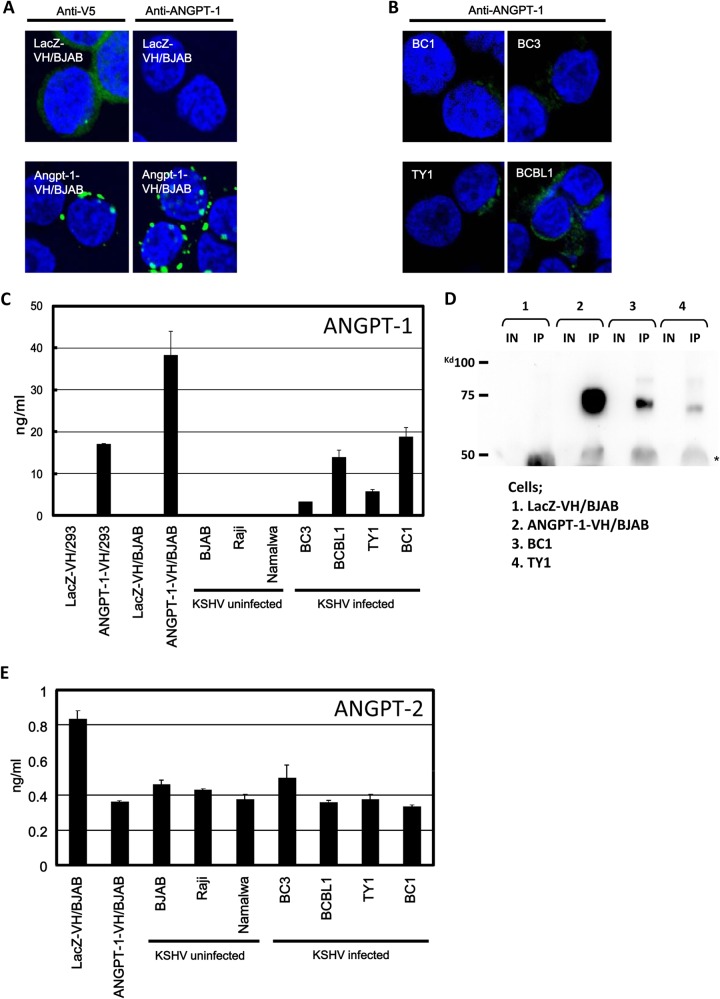
FIG 1.
ANGPT-1 expression in KSHV-infected PEL cells and ANGPT-1 and ANGPT-2 secretion into medium. (A and B) ANGPT-1-VH/BJAB and LacZ-VH/BJAB cells were stained with an anti-V5 antibody (A, left, green staining) and an anti-ANGPT-1 antibody (A, right, green staining), followed by anti-mouse IgG conjugated with Alexa 488. KSHV-infected BC1, BC3, BCBL1, and TY1 cells were stained with an anti-ANGPT-1 antibody, followed by anti-mouse IgG conjugated with Alexa 488. (C) ELISA of ANGPT-1 secreted into medium. KSHV-infected BC1, BC3, BCBL1, and TY1 cells and KSHV-negative BJAB, Raji, and Namalwa cells were cultured until they reached a density of 106/ml, and then the supernatant was harvested and ANGPT-1 was measured by ELISA (28). ANGPT-1-VH/BJAB and ANGPT-1-VH/293 cells were used as positive controls, and LacZ-VH/BJAB and LacZ-VH/293 cells were used as negative controls. (D) Detection of secreted ANGPT-1 by Western blotting. ANGPT-1 was immunoprecipitated from the supernatant with an anti-ANGPT-1 antibody and protein G Sepharose and subjected to Western blotting. IN, input; IP, immunoprecipitated. 1, LacZ-VH/BJAB; 2, ANGPT-1-VH/BJAB; 3, BC1; 4, TY1. (E) Detection of secreted ANGPT-2 with an ELISA kit. KSHV-infected PEL cells and EBV-infected or uninfected BL cells were tested as described for ANGPT-1 in panel C.

