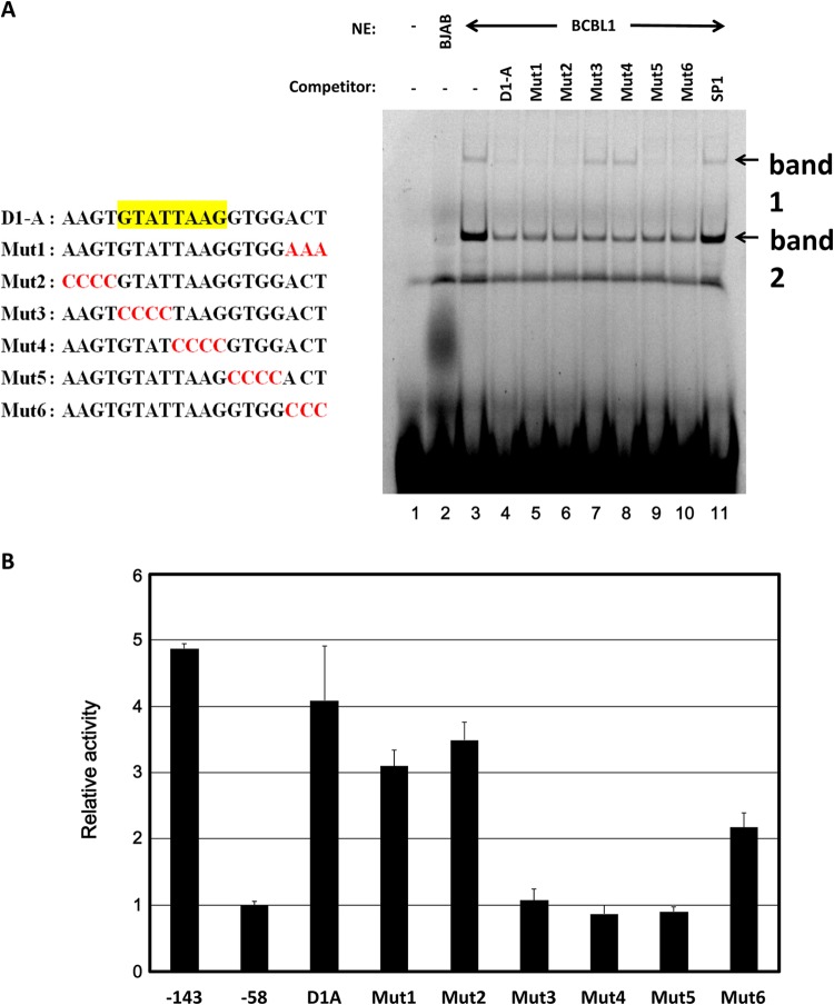
FIG 5.
EMSA of the D1A element. (A, left) Schematic illustration of the D1A region and mutant forms of the D1A region. (A, right) Nuclear extracts (NE) of KSHV-infected PEL (BCBL1) cells were mixed with D1A labeled with Cy5. A 20-fold concentration of each unlabeled mutant probe was used for the competition experiment. A minus sign indicates no nuclear extract or no competitor, respectively. In lane 2, 10 μg of BJAB nuclear extract was the input; in lanes 3 to 11, 10 μg of BCBL1 nuclear extract was used. (B) Mutant plasmids were constructed by inserting each mutant element into the region upstream of the bp −58 to +490 reporter, which contains the ANGPT-1 promoter. Data are shown as fold activity differences from the activity of the basal reporter; the activity of a bp −58 construct was set at 1-fold.

