FIG 3.
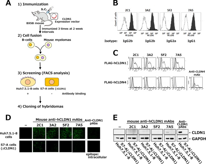
Establishment of mouse anti-hCLDN1 MAbs. (A) Strategy for MAb development against extracellular domains of hCLDN1. s.c., subcutaneous. (B) Huh7.5.1-8 (black histograms) or S7-A (white histograms) cells were incubated with the conditioned medium from each hybridoma clone and treated with phycoerythrin-conjugated secondary Abs. Stained cells were analyzed by flow cytometry. (C) 293T cells were transiently transfected with pcDNA3.1(+)-FLAG-hCLDN1 (FLAG-hCLDN1) or pcDNA3.1(+)-FLAG-hCLDN4 (FLAG-hCLDN4). After 2 days, cells were stained with anti-hCLDN1 MAbs or an anti-CLDN4 MAb and analyzed by flow cytometry. Gray and white patterns are for vehicle- and MAb-treated cells, respectively. (D) Huh7.5.1-8 (upper) and S7-A (lower) cells were incubated with anti-hCLDN1 MAbs or polyclonal Abs (pAbs) that recognized the cytosolic domain of CLDN1 and then treated with Alexa 488-conjugated secondary Abs. Stained cells were observed by confocal microscopy. (E) Huh7.5.1-8 or S7-A cell lysates were subjected to immunoblotting with anti-hCLDN1 MAbs, anti-CLDN1 pAbs, or an anti-GAPDH MAb.

