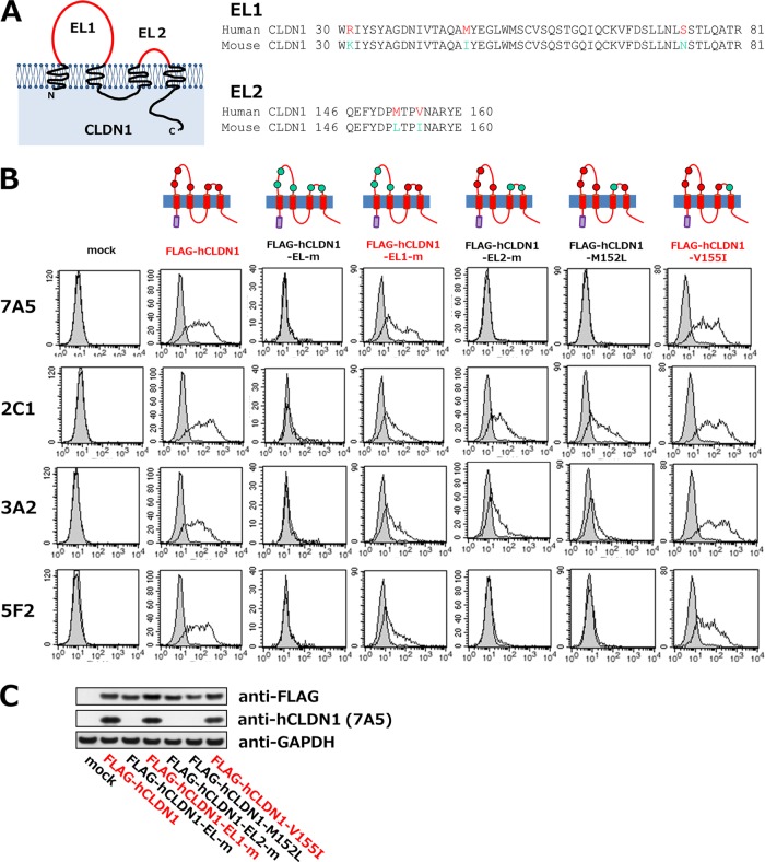
FIG 5.
Analysis of anti-hCLDN1 epitope on hCLDN1. (A) Schematic structure of CLDN1 and homologies of the first (EL1) and second (EL2) extracellular loops of human and mouse CLDN1 proteins. (B and C) 293T cells were transiently transfected without a vector (mock) or with the FLAG-hCLDN1, FLAG-hCLDN1-EL-m, FLAG-hCLDN1-EL1-m, FLAG-hCLDN1-EL2-m, FLAG-hCLDN1-M152L, or FLAG-hCLDN1-V155I expression vector, as indicated. (B) Flow cytometric analyses were performed using anti-hCLDN1 MAbs (7A5, 2C1, 3A2, and 5F2). Each cell culture was incubated with 2 μg/ml of anti-hCLDN1 MAb (white histograms) or control IgG (gray histograms), and Ab binding was detected using Alexa 488-conjugated goat anti-mouse IgG(H+L). (C) The cell lysates were subjected to immunoblotting with anti-FLAG, anti-hCLDN1 (7A5), and anti-GAPDH.

