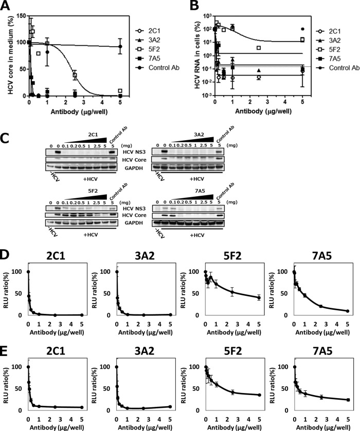
FIG 7.
Inhibition of HCV infection by anti-hCLDN1 MAbs in vitro. Huh7.5.1-8 cells were seeded at 5 × 10⁴ cells in 48-well plates and pretreated with the indicated amount of anti-hCLDN1 MAb or control IgG (control Ab) for 1 h at room temperature and then were subjected to the following procedures. (A to C) Cells were infected with HCVcc (MOI = 1) for 2 h at room temperature and cultured for 4 days in the presence of the indicated amounts (0.1 to 5 μg/well) of MAbs (400 μl/well). (A) Culture supernatants were collected, and levels of HCV core proteins were quantified by ELISA. (B) The cellular total RNA was extracted, and HCV RNA contents were quantified by qRT-PCR. The dotted line represents the average level in HCV-uninfected cells. In panels A and B, values are expressed as percentages, and data represent the means ± standard deviations (SD) (n = 3). (C) Cell lysates were subjected to immunoblotting of HCV NS3, core, and GAPDH proteins. (D and E) Cells were then infected with HCVpp (genotype 2a [D] or genotype 1b [E]) for 3 h at room temperature and cultured for 2 days in the presence of the indicated amounts of each anti-hCLDN1 MAb. Luciferase activities in cell lysates were measured using a luminometer. Values are expressed as percentages. Data in each graph represent the means ± SD (n = 3). RLU, relative light units.

