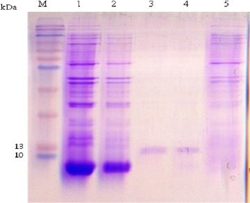
Fig. 3:

SDS–PAGE analysis of the rEPC1. The gel was Coomassie brilliant blue-stained., the expected molecular mass of the His6-tagged rEPC1protein was 12.8 kDa. M, protein marker; lane 1: Escherichia coli lysates with IPTG 0.5 mM after 6 h of induction, lane 2: Escherichia coli lysates with IPTG 1 mM after 6 h of induction, Lane 3 and 4: rEPC1 protein extracts, lane 5: Escherichia coli lysates without IPTG induction
