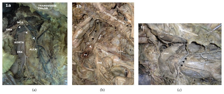
Figure 1.
Photograph of the posterior abdominal wall showing the aorta and inferior mesenteric artery (IMA) and its branches. (a) A thin arterial channel (arrowheads) connecting middle colic (MCA) branch of superior mesenteric artery (SMA) to the ascending branch of left colic artery (ALCA) is observed. (b) Arrowheads show an arterial connection between the celiac artery (CT) and the left colic artery (LCA) ascending branch. SplA: splenic artery, PV: portal vein, Trv colon: transverse colon. (c) An inner arterial channel (arrowheads) besides the marginal artery (Ma) connecting the proximal SMA to LCA. PANC: pancreas.

