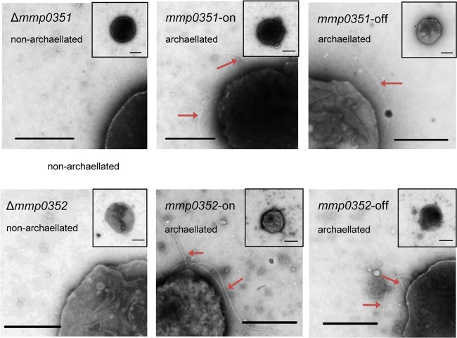
FIG 4.
Electron micrographs of mmp0351 (A) and mmp0352 (B) deletion mutants complemented with the wild-type version of the respective gene. The deletions alone are shown in the left panel followed by the complement-on cells grown in N-free medium with the addition of alanine and complement-off cells grown in N-free medium with the addition of NH4Cl. All samples were negatively stained with 2% phosphotungstic acid (pH 7.0). Arrows indicate archaella. Scale bars represent 0.5 μm.

