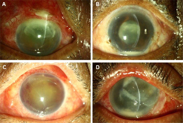
Figure 2.
Preoperative clinical photographs of a few exogenous filamentous fungal endophthalmitis cases.
Notes: (A), post cataract surgery endophthalmitis with corneal and scleral tunnel fungal infiltrate. (B), cobweb-like exudates in pupillary area and on intraocular lens. (C), organized coagulum in the anterior chamber. (D), post-pterygium excision keratitis and endophthalmitis.

