Figure 3.

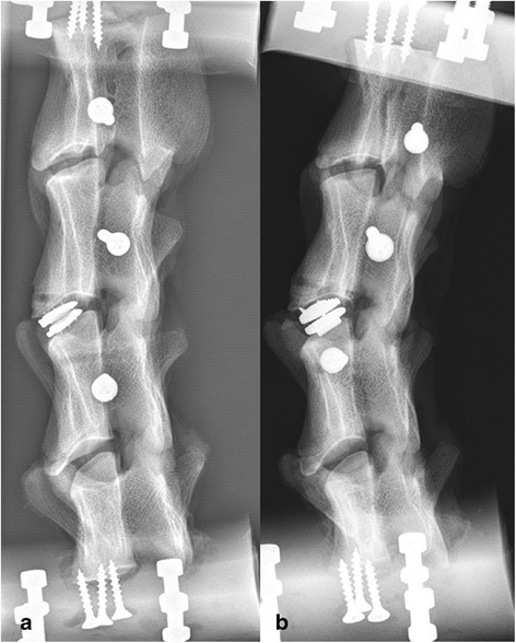
Lateral radiograph of two different specimens. Demonstration of the correct position of the Type D-prosthesis (a) and the Type A-prosthesis (b).

Lateral radiograph of two different specimens. Demonstration of the correct position of the Type D-prosthesis (a) and the Type A-prosthesis (b).