
Figures 3.
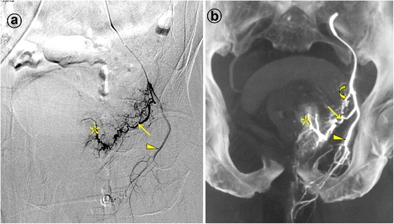
Prostatic artery arise from the internal pudendal artery. Images from a patient with severe lower urinary tract symptoms due to benign prostatic hyperplasia (117 mL) underwent PAE. a. Digital subtraction angiography (DSA) of the anterior division of the left internal iliac artery with ipsilateral oblique view demonstrates the left prostatic artery (straight arrow) and the left internal pudendal artery (arrowhead). The asterisk indicates the contrast staining in the left prostate lobe. b. Cone-beam CT image with coronal view after selective catheterization of the anterior division of the left internal iliac artery demonstrates the left prostatic artery (straight arrow) and the left internal pudendal artery (arrowhead). The curved arrow indicates the inferior vesical artery, which is difficult to identifying on the DSA. The asterisk indicates the contrast staining in the left prostate lobe.