Figure 4.

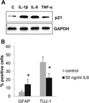
Proinflammatory cytokines induce p21 and decrease neurogenesis in NPC. (A) Western blot analysis of NPC differentiated in the presence of 10 ng/ml IL-1β or 50 ng/ml IL-6 or 20 ng/ml TNF-α for 8 days. (B) The graph depicts the percentage of Tuj-1+ neuroblasts and GFAP+ glia cells among NPC cells differentiated untreated (control) or in the presence of 50 ng/ml IL-6. Data are presented as a mean ± SEM, *p < 0.05. GAPDH, glyceraldehyde 3-phosphate dehydrogenase; GFAP, glial fibrillary acidic protein; Tuj-1, neuron-specific class III beta-tubulin.
