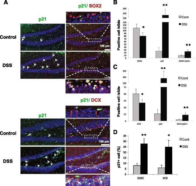
Figure 5.

Chronic intestinal inflammation induces SGZ p21. (A) The confocal microscopic analysis shows that p21 (nuclear, green) is co-localized with SOX2 (nuclear, pink) or DCX (cytoplasmic, red) and is more abundant in the SGZ of DSS-treated hippocampus. Five slides/group were analyzed and representative images are shown; arrows indicate cell expressing p21 and co-localization of p21 with SOX2 or DCX. (B) Average number of SOX2+, p21+, and SOX2+/p21+ cells/per slide. (C) Average number of DCX+, p21+, and DCX+/p21+ per slide. (D) Percent of p21+ cell in SOX2+ or DCX+ cells/ per slide in the hippocampus of control and DSS-treated mice. Six slides/mouse/five mice were analyzed. Data are presented as a mean ± SEM. Statistical analysis was performed with the Student t-test with Satterthwaite correction. *p < 0.05; **p < 0.01. Cont, control; DCX, doublecortin; DSS, dextran sodium sulfate; SOX2, sex-determining region Y-box 2.
