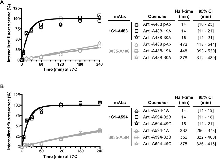
Fig 4. Internalization measurements of the anti-EphA2 mAbs with the anti-Alexa Fluor mAbs.
Cellular uptake of 1C1-A488 or 3035-A488 (A) and 1C1-A594 or 3035-A594 (B) over a 4-hr time course. Upon labeling with the antibody conjugate, PC-3 cells were pulsed at 37°C for up to 4-hr. Surface fluorescence was subsequently quenched with 1 of 2 anti-A488 mAbs (A) or 1 of 3 anti-A594 mAbs (B). A rabbit anti-A488 polyclonal was used as a benchmark for the A488 conjugated mAbs. Percent internalization was calculated as described in the Material and Methods section. One representative experiment of multiple is shown. An independent experiment for the anti-A488 antibodies is shown in S1 Fig.

