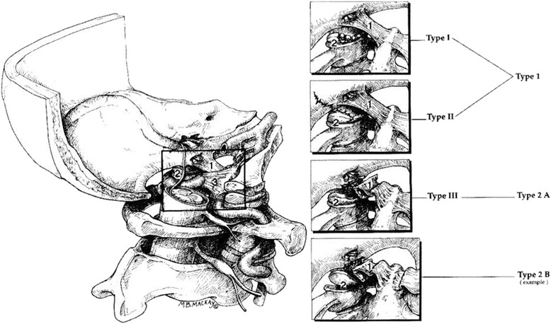
Figure 4.

Types of OCF based on the Anderson and Montesano classification system (Types I-III) compared with the Tuli classification system (Types 1, 2A, and 2B), it shows the left craniocervical junction from its medial aspect. The dura and the inferior aspect of the alar ligament have been removed to show the fractured condyles in the fracture types. Tuli S, Tator C.H, Fehlings M.G, Mackay M (1997) Occipital condyle fractures. Neurosurgery;41:368-76.
