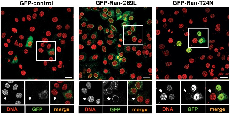
Fig 2. Inter-cellular transfer of Ran.
HeLa cells were transfected with indicated constructs for 9 h and were then co-cultured with untransfected NIH3T3 cells for 18 h. Cells were stained with GFP antibody (green) and the DNA dye Hoechst 33342 (pseudocoloured in red). Arrows indicate NIH3T3 cells as detected by the characteristic punctate staining of the nucleus. Scale bar, 25 μm.

