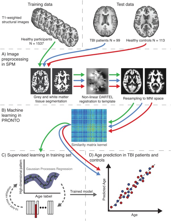
Figure 2.
Overview of the study methods. Study data comprised 2 sets, training and test. The training set used structural magnetic resonance imaging from 1,537 healthy individuals from multiple cohorts, whereas the test set included 2 groups, 99 traumatic brain injury (TBI) patients and 113 healthy controls, all scanned on the same scanner. (A) Conventional Statistical Parametric Mapping (SPM) structural preprocessing pipeline was used to generate gray and white matter maps, normalized to Montreal Neurological Institute (MNI) space and modulated to retain data relating to brain size. (B) Separately for gray and white matter, all 1,749 data sets were converted to a kernel matrix based on voxelwise similarity using Pronto. (C) The training data only were run through a supervised learning stage where a Gaussian Processes Regression (GPR) machine was trained to recognize patterns of imaging data that matched a given age label. To assess model accuracy, 10-fold cross-validation was conducted where 10% of samples were excluded from the training step and the ages of these samples were estimated. This was iterated 9 further times to generate age predictions on all samples. (D) The trained GPR model was then applied to the 2 test data sets, to assess accuracy of the model on healthy controls and then predict brain age of TBI patients. [Color figure can be viewed in the online issue, which is available at http://wileyonlinelibrary.com.]

