
Figure 2.
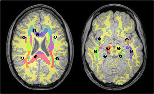
Measuring FA in white matter areas. The segmented white matter mask (yellow) is shown overlaid on a high-resolution T1-weighted image registered to standard MNI space. The ICBM-DTI-81 atlas (coloured areas) was used in order to measure the FA in areas defined as white matter according to the segmented mask in specific tracts: 1, genu of the corpus callosum; 2, body of the corpus callosum; 3, splenium of the corpus callosum; 4, superior longitudinal fasciculus; 5, cerebral peduncle; 6, sagittal stratum; 7, cingulum; 8, uncinate fasciculus.