
Figure 3.
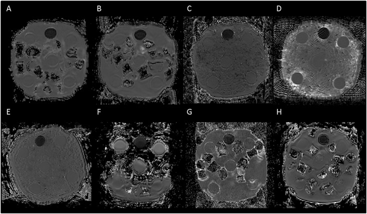
Comparison of the ice–water phantom ADC images across scanners. Images of the phantom from each of the scanners using the same contrast range are shown. The tube filled with sucrose appears darker, while the five tubes filled with distilled water are evenly separated from each other and can be seen to be surrounded by ice–water. The ice–water was prepared by using either ice cubes (e.g. scanner A) or crushed ice (e.g. scanner C). The protocol for scanning the phantom was not adhered to for scanner F, with the image showing that ice–water was not surrounding all of the tubes at acquisition.